How to Manage Hip Arthritis

Osteoarthritis

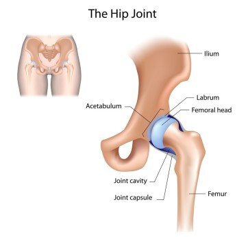
Osteoarthritis (OA) is the most common cause of hip pain in adults over 50. Simply put, OA is a degeneration of the hip joint. The cartilage that lines the joint degenerates over time until the femur (ball) and the acetabulum (socket) no longer have the lubrication and protection required for normal, smooth joint motion.  This can become quite painful and ultimately lead to decreased activity levels and increased difficulty with daily tasks such as walking and standing up, especially first thing in the morning.

Treatment for hip OA can include stretching and strengthening of the muscles surrounding the hip. Ultrasound and hot packs can be used in the short-term management of pain and activity limitations in order to return to exercise and begin therapy quicker. Manual therapy including joint mobilization, soft tissue release and massage all have significant evidence supporting their use to improve function and reduce activity limitations in people with OA.

2017 updated risk factors for osteoarthritis of the hip include, but are not limited to:

  • Decreased hip internal rotation and flexion range of motion
  • Morning stiffness
  • Male sex
  • Higher BMI
  • Hip pain
  • Age over 50

Hip Internal Rotation : range of motion becomes limited


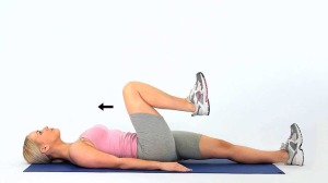
Hip Flexion also becomes restricted and painful


We’re here to help. In many cases, a prescription of manual therapy to improve hip range of motion, as well as an individualized exercise program can help to prolong or prevent joint replacement surgery.

Treatment recommendations include:

  • Ultrasound for short-term pain reduction
  • Flexibility, strengthening and endurance exercises
  • Manual therapy for joint mobility, flexibility and pain
  • Education on activity modification, exercise, supporting weight reduction when overweight, and methods of unloading arthritic joints.

Recent evidence suggests that NSAIDs (such as Aspirin and ibuprofen), COX-2 inhibitors, and steroid injections are effective at relieving pain and symptoms in hip OA. However, there is some evidence that suggests NSAIDs may increase the progression of hip OA my decreasing glycosaminoglycan synthesis. In addition, there can be serious gastrointestinal side effects associated with NSAID use. Consult your physician before use.

What can you do at home?

Below is a video with a few stretches recommended to improve hip mobility. This is not an exhaustive list but rather a starting point and something to try. It is always recommended that you see a physical therapist to individualize your treatment approach.

The information provided is not medical advice. If you would like more information please visit us at www.ActiveMarin.com or call to schedule an appointment. 

Achilles Tendon Tears: Are you at Risk?

The Achilles tendon is the largest and strongest tendon in the body, but unfortunately the aging tendon undergoes various changes that put it at risk for injury. Some changes that increase with age include decreased collagen density, decreased glycosaminoglycans and water content, decreased tensil strength and increased stiffness.

A study of 891 ruptured tendons in humans revealed 97% of the changes were degenerative in nature and about 50% of them were Achilles tendons.

How can you keep your Achilles tendons healthy?

Know your risk factors associated with Achilles tendon disorders and schedule an appointment for a physical therapy evaluation to determine the likelihood of your developing an Achilles tendon disorder and to learn research proven treatment strategies to help strengthen and avoid surgical repair.

Risk factors include:
-abnormal dorsiflexion (increased or decreased ability to flex foot)
-abnormal subtalar range of motion (increased or decrease foot joint mobility- pronation or supination)
-decreased plantar flexion strength
-abnormal tendon structure
-medical conditions associated with Achilles tendon disorders: diabetes, obesity, high blood pressure, high cholesterol

Treatment:
Studies have shown and proven that eccentric exercises with a focus on slow and controlled movement is an extremely effective nonsurgical method to treating Achilles tendonitis/tendonosis. Below are some helpful exercises for anyone dealing with symptoms or has been diagnosed with this very common injury.

Eccentric Heel Drop on Step: (Achilles Tendon Strengthening)
Begin this exercise with both feet in a neutral position with only the forefoot on the step
Perform a toe raise with both feet
Remove the unaffected leg from the step and slowly lower the affected back to neutral, keeping knee straight
Perform 3 x 15 repetitions twice a day. This is maintained every day for 12 weeks.
As soon as 2 x 15 repetitions twice a day can be done pain free, the load should be increased.
Progression:  Loaded Eccentric Heel Drop on Step–now add weighted dumbells to increase difficulty and to build strength

What Is The Difference Between Dizziness and Vertigo?

vertigo-treatment

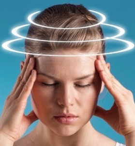
People often use the word “dizziness” when talking about a variety of symptoms. It is important for you   to be able to differentiate between these symptoms,  because the cause, diagnosis and treatment cn be quite different. Dizziness is a sensation of lightheadedness, faintness, or unsteadiness. Unlike dizziness, vertigo has a rotational, spinning component, and is the perception of movement, either of the self or surrounding objects.

Vertigo is often caused by an inner ear problem.  One common cause is called  benign paroxysmal positional vertigo (BPPV). This occurs when tiny particles of calcium form in canals of the inner ear. The inner ear sends signals to the brain about head and body movements relative to gravity. With this move of crystals from one area to another, the inner ear tells the brain you’re moving when you are really not.

Physical therapists can perform specific testes to determine if you have BPPV and will guide the treatment. One such test includes examination of the eyes for nystagmus, an involuntary rapid eye movement. If BPPV is found, the patient is guided through a repositioning treatment involving the head and neck called the Eppley maneuver. The PT will guide you through a series of position changes designed to relocate the crystals to their appropriate area in the inner ear. With treatment, symptoms should significantly decrease or completely resolve.

Physical therapy can play a strong role in recovery of symptoms. Treatment would involve an exercise-based program to improve balance and reduce symptoms related to dizziness. Specifically, treatment would include:

● visual stabilization training
● postural education
● balance training
● cervical mobility and stretching

Expected outcomes from treatment include decreased risk of falls, improvement in balance, improved ability to stabilize vision, an increase in cervical mobility, and a return to prior level of function.

Rotator Cuff Tears: Physical Therapy vs. Surgery, Is There A Difference?

The Rotator Cuff and Shoulder Girdle

By Megan Morgan, DPT

A recent study that was conducted in Finland determined that Physical Therapy alone for non-traumatic rotator cuff tears (supraspinatus) results in equal functional outcomes at one year compared to surgically repaired tears. All of the groups in the study received the same Physical Therapy (PT) instruction and prescription of a home exercise program as well as 10 PT sessions. The study found that patient’s who did receive surgery did not report any superior results than those who only received PT.

rotator-cuff-physical-therapy

Role of the Rotator Cuff and Scapular Stabilizers

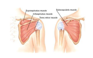
The rotator cuff is a group of four muscles that connect the humerus (upper arm) to the scapular (shoulder blade). These muscles not only create movement of the arm but they play an integral role in providing stability to the shoulder joint. The tendons of these muscles form together to create the cuff; which work together to create a force couple relationship that results in the humerus to remain centered on its base of the scapula (also known as glenoid). When a muscle of the rotator cuff is torn dysfunction of this force couple relationship results and the shoulder has difficulty maintaining its stability.

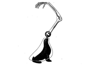
Another critical component of a healthy functioning shoulder is scapular strength and proper muscle activation. The scapula is the base that connects our humerus to our body via the placement of the humeral head on the glenoid of the scapula. There are multiple muscles that connect our scapula to our spine and these are referred to as our scapula stabilizers. A common analogy for the role of these stabilizers is a seal trying to balance a ball on its’ nose; they have the role of creating a stable base so the ball (humeral head) can stay balanced for the humerus to move on. These muscles are just as important to providing stability to the shoulder joint, as the rotator cuff. Therefore it is important to strengthen these stabilizers and create the proper awareness of one’s scapula position. Physical Therapists are here to help develop your awareness and the appropriate individualized strengthening program.

rotator-cuff-strengthening

The study was limited in that it only incorporated individuals whose tears were <75%. Therefore, we cannot apply these findings for all rotator cuff tears, as the severity of tears varies between individual cases. But an important take away message from this study is that for partial rotator cuff tears PT and a home exercise program can independently get you back to your prior activities of choice.

References:
https://www.apta.org/PTinMotion/NewsNow/2014/3/12/RotatorCuffTears/?blogid=10737418615